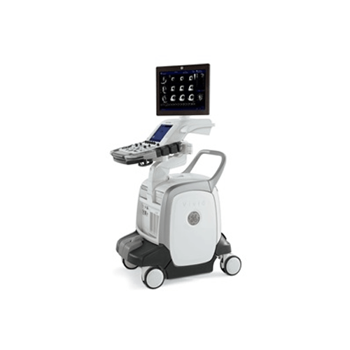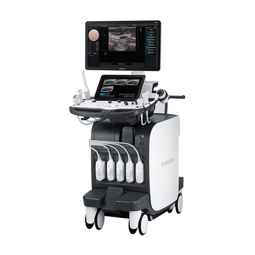
- 23" Full HD LED Display for Exceptional Image Contrast
- Ergonomic Design for Ease of Use
- Comprehensive selection of transducers available for a Wide Range of Clinical Applications
-

-
- Outstanding clinical performance to support confident diagnoses
- Extremely versatile with easy-to-use 3D/4D capability
- Designed for optimal workflow and reliability in today’s busy practices
- Access to Philips award-winning customer service and Remote Services Network
-
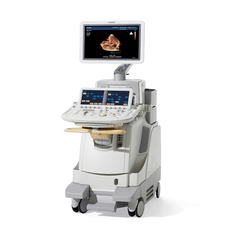
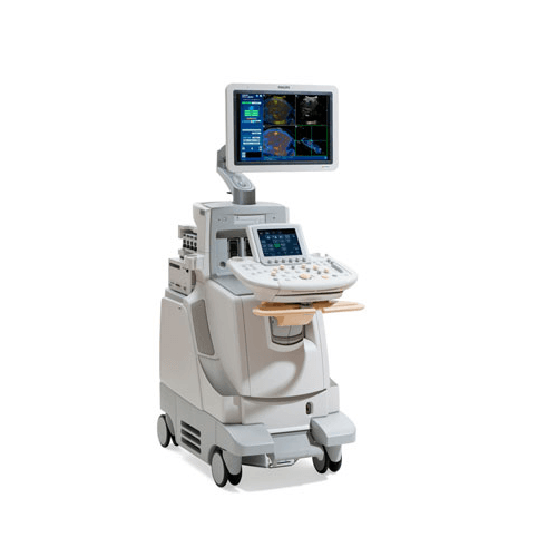
Combining unprecedented 2D and 3D image quality in the same transducer and a host of easy-to-use quantification, clinical performance, and information management tools, the new iE33 xMATRIX echo system addresses the clinical needs of managing patients with cardiac disease, including heart failure, valvular disease, and congenital heart disease.
-
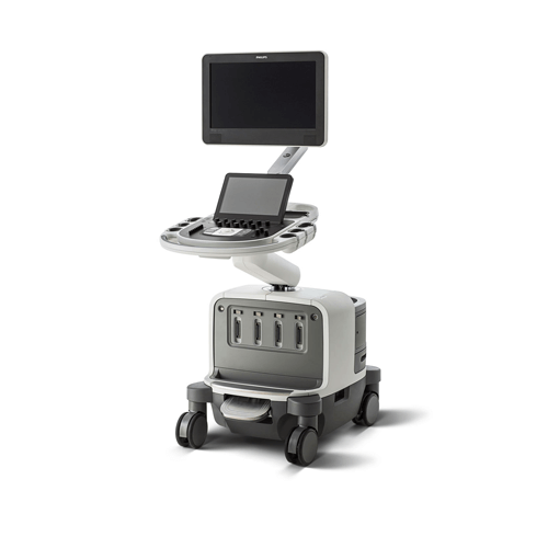
EPIQ 7 features our most powerful architecture ever applied to ultrasound imaging – touching all aspects of acoustic acquisition and processing, allowing you to truly experience ultrasound’s evolution to a more definitive modality. EPIQ 7 is supported by our family of proprietary xMATRIX transducers and our leading-edge Anatomical Intelligence.
-
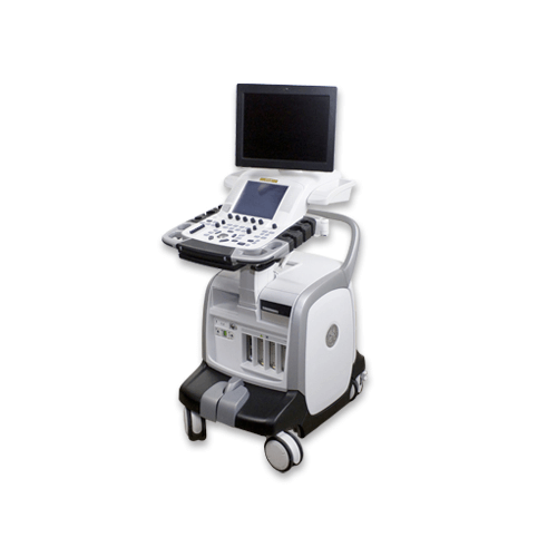
The Philips iU22 is a premium shared service ultrasound system that is the brother of the Cardiac focused iE33. Unlike the iE33, the iU22 focuses on General Imaging and OB/GYN applications, but still retains the same shared service functionality. Imaging and features are top of the line only recently replaced by the newer Epiq 5.
-
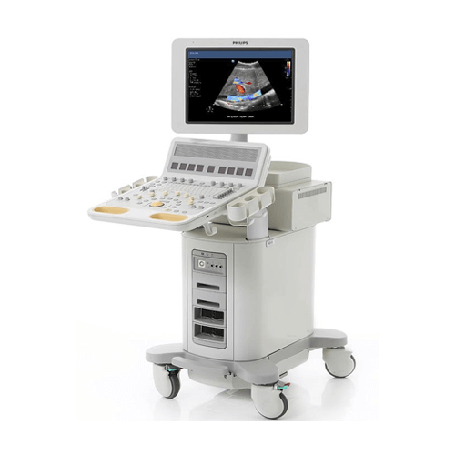
- CrossXBeam
- 3D/4D Volume obstetric imaging
- 2D/3D/4D modes
- PW/Color/Power Doppler (CW Doppler as option)
- Anatomical M-Mode
- ECG module with CW upgrade
- SRI HD High Definition Speckle Reduction Imaging
- 19″ LCD monitor
- Report Writer
- Integrated DVD Multi Drive
- Height and rotation adjustable keyboard
- Backlit Keyboard
- Integrated Gel Warmer
- B-Flow
- B-Steer+ Needle Visualization
- Stress Echo Option
- Lightweight
- 7-inch LCD touchscreen interface
- Elastography Option and Elastography Quantification option
- Contrast enhanced ultrasound
- Export to USB memory stick or CD/DVD
- DICOM
- TVI Tissue Velocity Imaging
- Cine Ultrasound Review
-
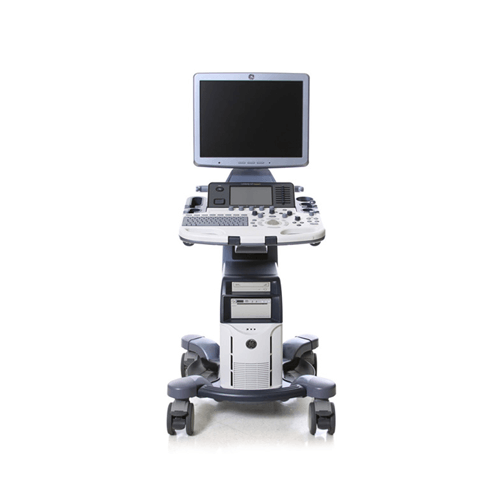
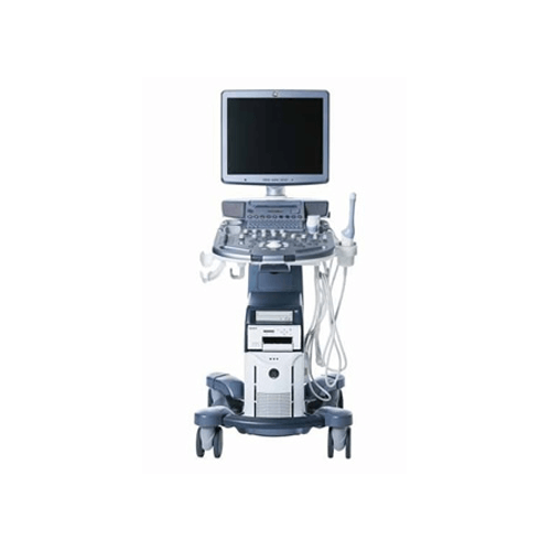
The Voluson S8 system opens new opportunities in clinical imaging while enabling the efficiency and productivity that busy practices demand. Among its advantages:

- Extraordinary image quality — the foundation of Voluson ultrasound— to help achieve a clear view into obstetric and gynecologic exams.
- Sophisticated fetal assessment tools — to help support earlier, more detailed evaluations.
- Innovative probe technologies — to support thorough evaluations of even the most challenging patients.
- Easy imaging — with system intelligence and probe technology combining to produce outstanding images with minimal user interaction.
- Easy-to-use automation tools — that help streamline workflow, forge stronger connections with patients and referring physicians, and control costs.
- Ergonomic design — that simplifies how users interact with the system and helps provide optimal comfort while scanning.
-
- CrossXBeam
- Integrated DVD
- HD Speckle Reduction
- Integrated Gel Warmer
- 2D, M-Mode
- PW/Color/Power Doppler
- B-Flow
- B-Steer+
- Flow Quantification
- Stress Echo Templates
- Stress echo wall motion
- Lightweight
- 10-inch LCD touchscreen interface
-
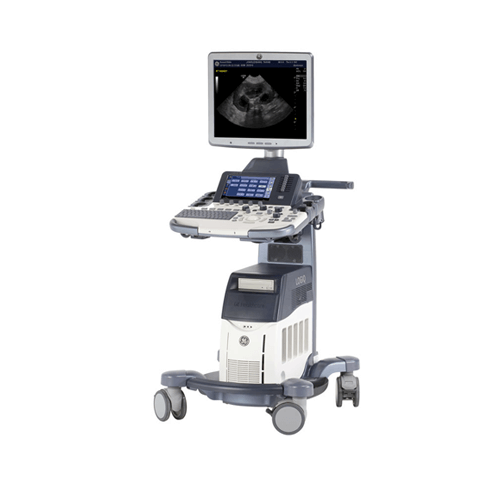
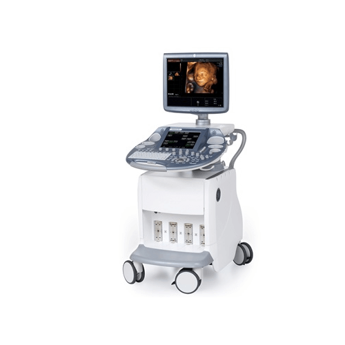
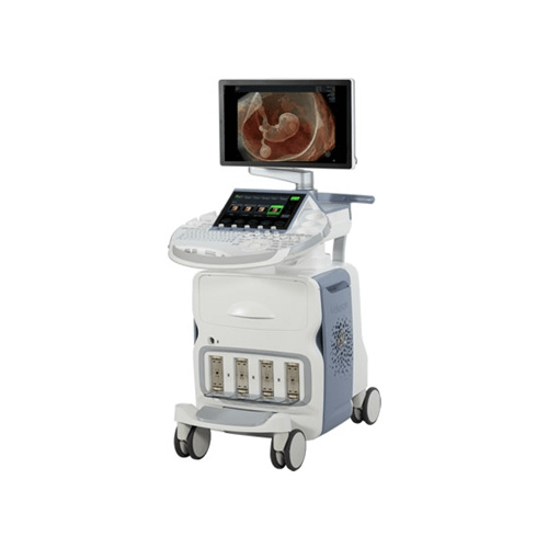
The powerful GE Voluson E6 is designed specifically to give you the exceptional vision you need today so you have the flexibility to meet your emerging needs. This versatile system helps deliver expandable capabilities to grow with your practice. Voluson E6 has enhancements to help you improve patient care and simplify your workflow.
-
- Explain current and advanced ultrasound technologies
- Describe system physics and instrumentation
- Understand how 2D strain/speckle tracking works and how strain data is acquired and displayed
- Demonstrate basic and advanced image optimization, customization and data management principles
- Understand 3D/4D image acquisition, visualization and quantification tools
- Identify areas you can incorporate advanced features into your workflow
-
- Deliver extraordinary image quality on a broad spectrum of patient body types.
- Visualize blood flow without the limitations of Doppler
- Enhance your workflow.
- Integrate real-time ultrasound with previously acquired CT, MR, PET, or ultrasound images.
- Visually track your position during a scan.
-
- 4x the processing power of the Voluson e10
- 4D Electronic Matrix 4D Transducer
- HD Live
- V-SRi advanced volumetric speckle reduction
- 4D Electronic Matrix 4D Transducer
- 23” Widescreen LED monitor
- 12” multitouch touch panel
- Probe port illumination
- High frame rates in 2D/4D imaging
- Ergonomically designed for maximum comfort
-
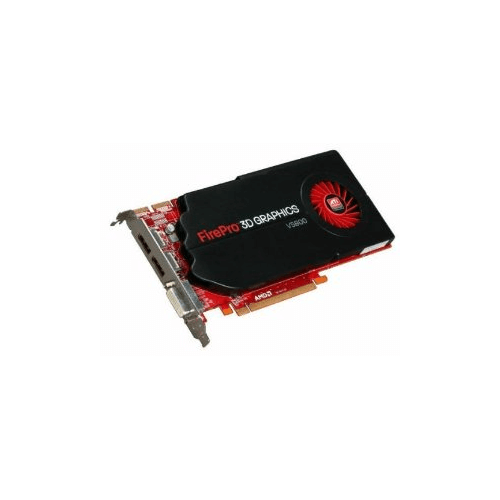
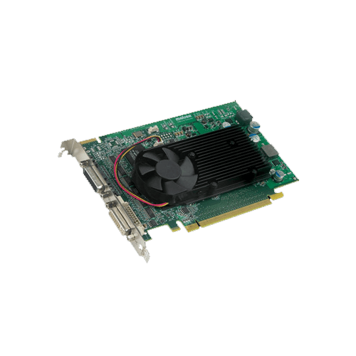
- Outstanding performance for the 3D mid-range segment
- 2.5X computing capability over prior generation1
- AMD Eyefinity multi-display technology2
- Drive up to 3 independent 30’’ displays3 12.3 million pixels4
- Scalable ultra parallel processing architecture with 800 stream processors
-
- PCIe x16 single-slot board with triple high resolution digital-display output
- DICOM compliant 8/10/13-bit LUT GAMMA ramp correction supported on all outputs in independent and/or stretched (NT-style) desktop modes for accurate display calibration
- Digital Luminance Correction™ (DLC™)1–enables calibration packages to uniformly calibrate display luminance to well-within industry standards.
- Dynamic Field-of-View Correction (DFC)1–with appropriate 3rd party applications, DFC reduces color and/or grayscale inaccuracies caused by different viewing angles of LCDs.
- Image Color Profiling (ICP)1–allows 3rd party developers to use hardware-enabled ICP with up to 13-bits for accurate color rendering of subtle anatomical image structures.
- Programmable Gamma LUTs supporting 8, 10 and 13-bit wide formats–for the highest calibration, meeting or exceeding DICOM industry standards across the entire display.
- Hardware Window IDs and LUTs–with multiple screen regions available, application can exploit hardware-accelerated window and level functionality on a specified region.
- Hardware pivot and cursor support as well as optimized bus performance and increased memory provide fast and smooth user experience for window & level and cineloops.
- Enhanced features available through Matrox imaging Library (MIL) to enable hardware-accelerated operations such as image cached window/levelling, zoom, and the ability to view 1024 simultaneous shades of gray from 16-bit source images.
-
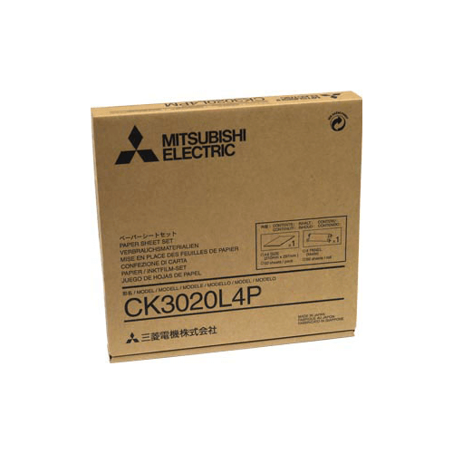
- 8x10 Matt Photo Paper and Dye Film
- 50 prints
- Printable area 8" x 10"
- Compatible printer - CP3020DAE.
-
- 8x10 Gloss Photo Paper and Dye Film
- 50 prints
- Printable area 8" x 10"
- Compatible printer - CP3020DAE.